Electromyography (EMG)
- Definition
- Description
- Preparation
- Risks
- Results
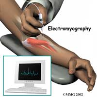
Electromyography (EMG) is a diagnostic procedure to assess the health of muscles and the nerve cells that control them (motor neurons).
An EMG uses tiny devices called electrodes to transmit or detect electrical signals.
During a needle EMG, a needle electrode inserted directly into a muscle records the electrical activity in that muscle.
The neurologist may insert needle electrodes at different sites depending on your symptoms.
After the EMG you may experience some temporary, minor bruising where the needle electrode was inserted into your muscle. This bruising should fade within several days. If it persists, contact your primary care doctor.
Take a shower or bath shortly before your exam in order to remove oils from your skin. Don't apply lotions or creams before the exam.
EMG is a low-risk procedure, and complications are rare. There's a small risk of bleeding, infection and nerve injury where a needle electrode is inserted.
EMG results can reveal nerve dysfunction, muscle dysfunction or problems with nerve-to-muscle signal transmission.
The neurologist will interpret the results of your exam and prepare a report. Your primary care doctor, or the doctor who ordered the EMG, will discuss the report with you at a follow-up appointment